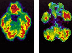
Any reader who was compos mentis during the Reagan-era War on Drugs will remember this ad. A man holds up an uncooked egg and says, “This is your brain.” Then he cracks open the egg and drops it onto a hot iron skillet. The egg sizzles and solidifies, and the man says, “This is your brain on drugs.”
Today this dramatic public service announcement seems quaint. Instead of fried eggs, we are bombarded with brightly colored brain-shaped images produced by a bewildering array of scanning technologies that purport to teach us about biological correlates of behavior. Some of the images compare addicts and non-addicts. Others juxtapose the brains of murderers and non-offenders, evoking ideas of inborn criminality championed in the late nineteenth and early twentieth centuries by Cesare Lombroso. Still others offer “wiring diagrams,” which supposedly show just how much male and female brains differ.
Brain images are ubiquitous and compelling. Findings reported in prestigious journals often lead to weeks of press hype. They can sway a jury toward conviction or away from the death penalty, guide medical treatment, or influence schools to develop separate but unequal education for boys and girls. For instance, the ACLU is suing the state of Florida for training teachers of boys to “engage students with debate and discussion” and teachers of girls “to create connections.”
But what do brain images really tell us? What critical questions can a layperson ask to avoid being sucked into a brain vortex or wowed by new words such as “connectome”? Can we really link specific brain structures to particular behaviors? Or do these fascinating new finds get a critical pass because they feed into ingrained preconceptions about biology as a root cause of all things evil or inevitable?
Oddly, I want to begin answering these questions by considering an important new study of fruit fly larvae. In a tour de force involving the automated analysis of almost 40,000 larvae, a research team led by neuroscientist Marta Zlatic identified twenty-nine worm behaviors including such showstoppers as wiggling to escape predators, crawling backward, and executing sequences of left and right turns. The scientists used specially engineered fly strains and, with a blue light, were able to selectively activate small groups of brain cells in individual larvae. A larval fly brain contains about 10,000 neurons, and Zlatic’s group activated up to fifteen neurons at a time in order to observe what behaviors, if any, resulted. Using automation and sophisticated mathematical analysis, they looked at the effects of groups of neurons that covered almost the entire brain.
Their findings tell us several important things about simple brain function. First, small numbers of activated neurons can induce a behavior, but there is serious redundancy: thirty to forty different groups may elicit the same behavior. Second, a given set of neurons may not always produce the same kind of behavior, even in the same brain: the researchers might activate a neural network that had previously paired with escape behavior yet find a different behavior expressed upon the second activation. The authors speculate that between-worm differences in development or experience, or even an individual worm’s personality or “state of mind” at the moment of stimulation, could account for this lack of correspondence between brain stimulation and behavior.
A third conclusion from all this maggot watching relates to the National Institutes of Health’s human connectome project, which hopes to determine the brain’s entire wiring diagram. By combining connectivity information with brain snapshots taken while subjects enact specific behaviors, researchers hope eventually to understand—much as Zlatic and colleagues have done with fly larvae—which human brain circuits are involved in which actions.
There are huge hurdles. The structural map a connectome provides and the neuron-activity map derived from neural imaging still need to be tied to specific behaviors via a third map—a neuron-behavior map. And herein lies the rub. Even for fly larvae, the latter map is not absolute. New experiences or lapses in bodily activity change brain wiring on long- and short-term time scales. So an individual’s connectome at one age may differ importantly from that at a different age. To predict future behavior, we need to know about an individual’s history and development.
With these principles in hand, let us circle back to criminals, sex differences, and addicts. Consider the case of Grady Nelson, a Florida man who was convicted of murdering his wife, Angelina Martinez. During the 2010 hearing to decide if Nelson should be sentenced to death, his lawyer showed the jury an image suggesting that Nelson had an abnormality in his left frontal lobe. At least two jurors were impressed by the evidence, shifting the voting balance toward life imprisonment. As much as I oppose the death penalty, the outcome raises some basic questions. Was the brain scan taken at the time of the murder, or, more likely, after years in jail? Could the brain deformations be linked to the murder? Scientifically, the introduction of this neural image was pretty lame, but the emotional impact was huge, and it carried the day for the defendant, who escaped execution.
Or what about the flurry of news stories this past December with headlines such as “Brain wiring in men, women could explain gender differences,” all reporting on a publication in the Proceedings of the National Academy of Sciences, which used neural imaging to produce average connectomes for brains of several hundred males and females.
Again, the images are compelling, but the science is not. First, neither the Proceedings article nor any other reputable research has tied specific wiring diagrams to variation in behaviors or cognitive skills. Just as with the fly larvae, the activity of differently wired networks can lead to the same behavior. Indeed, in an earlier study, the Proceedings researchers showed only small differences in the “big skills”—map reading, social cognition, spatial processing—that supposedly separate men from women. Second, the researchers do not assess the possibility that different experiences of gender might themselves produce differently wired brains. Did the young people in their samples play the same sports, have the same hobbies, wear the same type of clothing, or study the same subjects in high school?
As a final example, there have been some interesting “in the moment” imaging studies on the brains of people using drugs. These show us which parts of the brain see increased or suppressed neural activity while under the influence. What such studies do not tell us is whether or how regular drug use alters brain wiring or how changes in behavior or consciousness (hallucinations and the like) emerge from altered patterns of brain activity.
To understand neural images as more than just pretty pictures, we need to know something about the experiential history of the individuals under study. We need to know when a pattern of interest first appears and how stable it is. We need to remember both redundancy and lack of correspondence—activating different wiring patterns can produce the same behavior, while stimulating identical wiring patterns sometimes produces different behaviors. When comparing groups—males vs. females, law-abiders vs. criminals, addicts vs. non-addicts—we should recall that a “difference” is really a mean difference between groups that mostly overlap. Knowing about an average difference does not tell us about an individual’s characteristics or capabilities. Development and socially induced plasticity bar us from explaining, predicting, or excusing behavior. And, regardless of scientific findings, we need well-wrought theories of social context and morality.
Biologists are great. I love ’em. Some of my best friends are. . . . But we cannot properly use a science of behavior to make critical decisions about justice and human potential without also calling on the wisdom and insights of philosophers, historians, theologians, artists, sociologists, and many others.
